For my scar I choose to do one after an ACL surgery. As told in the background story my character suffered from a torn cruciate ligament and meniscus. As the anterior cruciate ligament was torn it needed surgery. So of course I will look into the surgery procedures deeply to understand what is actually happening even though I kind of know as I waited for my sister when she had her surgeries. But because this blog is not in German it was even more important to do proper research on it so I can explain in properly. But it is not only important to know what happens during the surgery you also need to know the anatomy of the knee as the scar needs to be placed right at the end. Also knowing the anatomy it will also helps understand what parts will move later which is important should the scar be for something other than doing a photoshoot where the model sits still. You need to know how movement will affect your application.
Anatomy of the knee
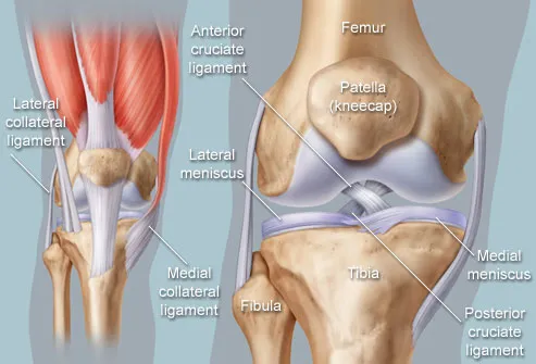
I found some good images which portray the knees anatomy quite well.


I will also take these images with me when doing the assessment as it is good for referencing.
On this website you can play around and see the different layers from 360°. It is very interesting as you normally can't see it. Also it shows all the different layers. Just bones, the connective tissues, deep muscles, muscles and at last with skin over it.
The knee joint consists of Femur(thigh bone), patella, Tibia(shin bone) and fibula. The lateral and medial collateral ligaments connect the femur with the fibula and tibia. On the tibia you can find the lateral and medial meniscus. Inside of the joint are the anterior and posterior cruciate ligament which keeps the joints in place and prevents overbending or overtwisting. But in some extreme cases( sports, motorbike accidents,etc) those ligaments can tear apart. In comparison with tendons the cruciate ligaments are not able to regenerate themselves and that's why surgery is necessary.
I found some good videos which explain the procedure but I will also explain it myself. On of the videos is a live surgery video so please be warned. Just in case you are not comfortable with those kind of things. I found kind of cool^^
After the injury happened you will have a very swollen knee it it could also happen that there is a concentration of blood inside. Which will have to be absorbed right away. You will also notice a decrease on motion and will definitely be in a lot of pain. Before surgery can be done the doctors have to wait for the swelling to go down. I also read that you should do physiotherapy until you have your surgery but when I think about how much my sister was in pain and couldn't properly walk I doubt that it is possible or comfortable for the patient. When it is finally the day of surgery the surgeon will first do a arthroscopy to see what exactly is damaged, how severe the damage is and to inspect the rest of the joint as well. At this point it will also be decided from where the new tissue will be taken. There are different types ans also the tissue can come from your own body ,which is the called autograft or you can get some from a donor ,which is called allograft. There is also synthetic graft. This is mostly use for mass ligament replacements or if you don't have access to an allograft or autograft. Commonly the tissue that is taken is a strip of your patellar tendon, part of hamstring tendons or part of quadriceps tendon.
Patellar tendon: part which is running from the bottom of your patella to the top of the tibia
Hamstring tendons:run from back of the knee on the inner side all the way up to your thigh
Quadriceps tendon: attaches patella to quadriceps muscle-> this is the large muscle on the front of the thigh.
After the surgeon know what the situation is you will be prepared for the surgery or more commonly now is that they do right after the arthroscopy as you are under anesthetics already anyway and it saves you from having multiple surgeries.
What is an arthroscopy?
It is a keyhole surgery. The arthroscope is a flexible tube that acts as camera and light source. The image from inside will then be shown on a monitor so the surgeon can see everything clearly. There will be some other some cuts on the sides of the knee as well for other medical instruments and a tube which will absorb blood and wound fluids. When staying longer in the hospital after a surgery on knee or leg you will have the tube for blood and wound fluids inside you for a a day or two and will be then be pulled out which can be painful and the skin around already started healing. But it is important that no fluids(blood,wound fluids) are just freely being inside you .But as those kind of surgeries are now also done in outpatient departments where the patient can leave around an hour after surgery you don't have this tube in you for days anymore.First the torn fibers of the anterior cruciate ligament are being removed and the new tissue which is most commonly taken from the hamstring tendons will be prepared. The new tissue is then fixed to some string and some kind of anchor which will secure it into the femur. There will also be a screw for fixating the other and in the tibia bone. Now the surgeon will drill a hole into the femur right through the tibia so the new tissue can be placed where the original cruicate ligament would have been.
I have made some screenshots of one of the videos for better understanding.
Here you see that above the knee joint the arthroscope is being inserted and two more cuts are being made for other medical instruments and the tube for absorbing wound fluids and blood

The next two show how the drill is making a tunnel through the femur and tibia for inserting the new tissue.


New tissue either from hamstring tendons or patellar tendon is being attached to a flexible string.

Tissue is being pulled through tunnel between the joints where the original cruciate ligaments would be.

Anchor that secures it in the femur.

And a screw for securing in the tibia. The bone will heal around it and accepts it as its own part.

After some time new cruciate ligaments will grow around the new tissue forming a new anterior cruciate ligament.

Also when taking new tissue from the hamstring tendons the surgeons tend to take more as needed to make sure they have enough but also as the hamstring tendon is able to reproduce new tissue from time to time.
After the surgery you will be walking in crutches for at least 3 weeks. Many also become a knee cast to keep your knee stable . I know my sister even had a wheelchair for the first days as she was in a lot of pain. Just a few days after surgery you will immediately start with physiotherapy to help your muscles build up again so your muscles can secure your knee joint and keep the ligaments stable.
I think the time of recovery is different for each individual as everyone has a different healing ability.
In my sisters case it took month until she felt secure enough to walk without the cast and one time her ligaments teared apart while having physiotherapy so you should be careful and how should I say it listen to your body. Of course a physiotherapist knows what he is doing, or at least should, but you know your body best and if you feel uncomfortable or think it is too much pressure on the knee you should stop.
Some other very common knee injuries are:
Fractures
Dislocation (hmm, tell me about it-.-')
Posterior cruciate ligament injuries
Collateral ligament injuries
Torn meniscus
Torn tendons
No comments:
Post a Comment